【Education】
・Diagnosis of gastrointestinal cancers (in the esophagus, stomach, small intestine, and large intestine)
Following advancement of the video-endoscopy system, gastrointestinal cancer can be detected at its early stage. Department staffs use a high-resolution endoscope to examine patients and perform dye-spraying endoscopic and magnifying endoscopic observations for lesions as necessary. Thus, gastrointestinal cancer is characterized with high accuracy.
・Endoscopic ultrasonography
Ultrasonography is performed for the gastrointestinal tract using endoscopy. The depths of cancer invasion and submucosal tumors can be determined.
・Treatment for early gastrointestinal cancers
The Department provides radical treatments for gastrointestinal cancers that are considered not to have metastasized (superficial esophageal, early gastric, early small intestinal, and early colorectal cancers) using an endoscope. Department staffs also use magnifying endoscopic observation, image-enhanced endoscopy (IEE) such as narrow band imaging (NBI) and blue laser imaging (BLI), and endoscopic ultrasonography to perform preoperative diagnostics with high accuracy and positively administer endoscopic treatment based on an accurate diagnosis.
・Diagnosis of inflammatory bowel diseases
Inflammatory bowel diseases include infectious enteritis, drug-induced enteritis, ulcerative colitis, and Crohn’s disease. The causes of infectious enteritis and drug-induced enteritis have been elucidated, but those of ulcerative colitis and Crohn’s disease have not. Department staffs make advanced diagnoses for these diseases using magnifying endoscopic observation, image-enhanced endoscopy (NBI and BLI), and endoscopic ultrasonography.
・Diagnosis of reflux esophagitis
Reflux esophagitis may cause heartburn. Inflammation occurs when gastric acid regurgitates into the esophagus. If reflux esophagitis continues over the long term, the esophagus may become stenosed. Department staffs also perform functional examination such as internal pressure measurements.
・Diagnosis and treatment of a gastroduodenal ulcer
A gastroduodenal ulcer, which may cause hemorrhage and perforation, is cured by the administration of a gastric acid secretion inhibitor or removal of bacteria from the stomach (Helicobacter pylori). Endoscopic hemostasis is performed in cases of bleeding.
・Treatment of gastrointestinal stenosis
Gastrointestinal stenosis has various causes, such as cancer and cicatricial stricture after endoscopic treatment. Bougienage, balloon dilation, and stent placement are performed for gastrointestinal stenosis.
・Diagnosis and treatment of small intestine lesions/diseases
The entire small intestine can be examined using double-balloon enteroscopy. Department staffs perform endoscopic diagnoses of small intestine lesions/diseases and histological diagnoses using biopsy as well as balloon dilation, endoscopic hemostasis, and polypectomy.
・Capsule endoscopy
The small intestine can be examined using a capsule endoscopy (CE) with a width of 11 mm and length of 26 mm. The CE is swallowed with a glass of water after fasting, and it moves forward in the gastrointestinal tract because of peristalsis. No abnormal sensations, such as uneasiness, occur owing to passage of the CE. Therefore, the small intestine can be examined while a patient performs their daily activities.
In January 2014, the National Health Insurance scheme began offering coverage for expenses incurred in undergoing colonic CE. Department staffs can currently perform colon CE. However, the National Health Insurance scheme can only cover patients into whom an endoscope is difficult to insert during conventional colonoscopy.
・Other services
1.Gastrostomy
2.Hemostasis for gastrointestinal bleeding
3.Foreign body removal
4.Measurement of internal pressure of gastrointestinal tract
5.Diagnosis and treatment of esophagogastric varices
6.Endoscopic diagnosis and treatment for hepatopancreatic diseases
【Research】
(Clinical research)
1.Development of new diagnostic modality for gastrointestinal (GI) tumor (image enhanced endoscopy, magnifying endoscopy, etc).
2.Analysis on clinical usefulness of endoscopic ultrasound for GI tumor.
3.Development of diagnostic and therapeutic strategy using double-balloon endoscopy for small-bowel lesions.
4.Analysis on clinical usefulness of capsule endoscopy for GI tract (small-bowel, colon)
5.Analysis on clinical study of Helicobacter pylori associated GI tumor (gastric carcinoma, MALT lymphoma, etc.).
6. Development of new treatment for inflammatory bowel disease (IBD) patients.
7.New development of therapeutic endoscopy for GI tumor, including laparoscopy and endoscopy cooperative surgery (LECS).
8.New medical examination for early detection of gastric carcinoma using blood (pepsinogen I/II).
9.Medicine-engineering collaboration for the development of image diagnosis and endoscopic equipment.
(Basic research)
1.Molecular biology for carcinogenesis of gastrointestinal (GI) tumor.
2.Pharmacogenomics of drug for IBD patients.
3.Establishment of the expanded indication of endoscopic resection for GI tumor (esophagus, stomach and colon).
4.Pathologic oncology of GI tumor.
5.Carcinogenesis of Helicobacter pylori associated GI tumor (gastric carcinoma, MALT lymphoma, etc.).
6.Genomic analysis of early GI tumor and IBD.
(Multicenter study)
1.Clinical study of detectability for colorectal lesions using wide-angle colonoscopy.
2.Clinical usefulness of NICE classification with narrow band imaging (NBI) endoscopy for colorectal lesion.
3.Colon capsule endoscopy (detection, preparation).
4.Effectiveness and safety of colorectal endoscopic submucosal dissection (ESD).
5.Long-term prognosis after ESD for patients with GI tumor.
6.Long-term prognosis of T1 colorectal carcinoma.
7.Prevention of stenosis after ESD for esophageal carcinoma using steroid intake.
8.Clinical usefulness of prototype double balloon endoscopy for small-bowel.
9.Detection of colitic cancer/dysplasia using chromoendoscopy and NBI for ulcerative colitis.
10.Endoscopic resection of colorectal polyps in patients with familial adenomatous polyposis.
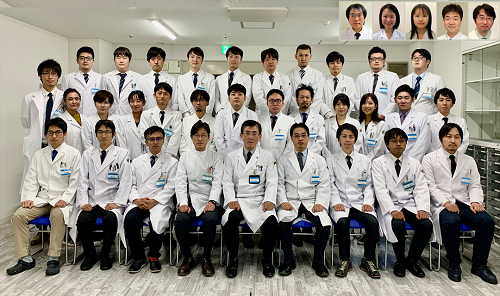
【Photo explanation】Staff of Endoscopy and Medicine
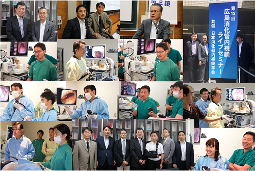
【Photo explanation】12th Hiroshima Gastrointestinal Endoscopy Live-seminar
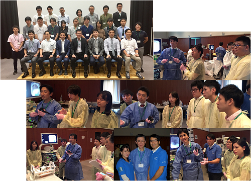
【Photo explanation】10th Hiroshima EMR/ESD Hands-on Seminar
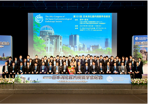
【Photo explanation】101st Congress of the Japan Gastroenterological Endoscopy Sosiety
【Photo explanation】76th Annual Meeting of Japan Society of Coloproctology

 Home
Home