
Masayuki Shimada, Ph.D., Professor
Graduate School of Biosphere Science, Hiroshima University
1-4-4 Kagamiyama, Higashi-Hiroshima City Hiroshima, Japan 739-8528
E-mail: mashimad * hiroshima-u.ac.jp (Please change * into @)
 Home
HomeFertility decreases slightly in women around the age of 30, but is clearly evident at a median age of 40. As a consequence, many women in developed countries including Japan, USA, UK and EU countries rely on assisted reproductive technology (ART) to become pregnant. However, the success rate of ART in women more than 40 years old women is low. Although a reduced ovarian reserve (number of oocytes recovered by controlled ovarian stimulation) may account for reduced success in some women using ART, the response of some older women to FSH is reduced and fewer follicles develop; these patients are called as “low responders”. The low responder patients have irregular menstrual cycles and an abnormal endocrine profile (high FSH / high LH / high androgen / low AMH). However, little is known about how the abnormal endocrine profile is induced or how it mediates a decline in ovarian function.
The collaboration research team, Hiroshima University and Baylor College of Medicine, revealed that the granulosa cells-specific Nrg1 knockout mice (gcNrg1KO) presented abnormal ovarian and endocrine phenotypes similar to those associated with reduced fertility in older women. Histological analysis of the ovary in 6-month-old gcNrg1KO revealed that follicular development was blocked in bi-layer secondary follicles and the ovarian stroma accumulated abnormal heterogeneous cells distinguished as two distinct types: LH receptor-positive endocrine cells and actin fiber-rich fibrotic cells expressing and accumulating collagen (Figure 1a,b). Both abnormal endocrine and fibrotic cells disappeared in response to long-term GnRH antagonist treatment (Figure 2), indicating that elevated LH and the abnormal endocrine functions induced the growth of fibrosis in the ovarian stroma. Follicular development to the antral stage was restored. Moreover, when the mutant mice and WT mice more than 15 months of age were treated with the GnRH-antagonist normal estrous cycles and pregnancies were restored with live pups delivered at least for 3 months (Figure 3).
This is the first report to document that the growth of fibrosis tissue in ovarian stroma is induced by the high serum levels of LH present with increasing age. The fibrosis is associated with the arrest of follicular development and the induction of abnormal endocrine profiles. Thus, GnRH antagonist treatments might provide a new, non-invasive strategy for improving fertility in a subset of non-responder aging women before menopause.
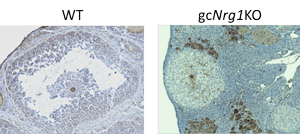
Figure 1a. The localization of LHR in ovaries of 6-months old WT and gcNrg1KO mice.
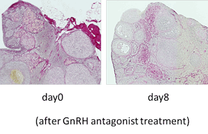
Figure 2. GnRH-antagonist treatment reduces the number of heterogeneous cells in ovarian stroma.
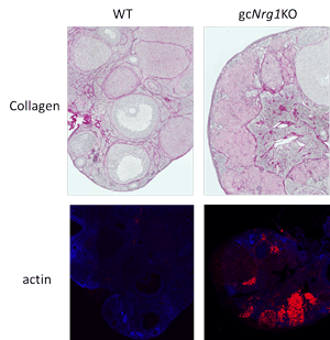
Figure 1b. The change of ovarian morphology with increasing is accelerated in gcNrg1KO mice.
Ovary collected from the day 2 after estrus was fixed by 4 % paraformaldehyde. The sections were stained by picrosirius red and eosin to observe collagen accumulation or rhodamine phalloidin, that is recognized F-actin, and DAPI, that is recognized nuclei.
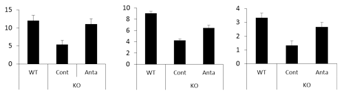
Figure 3. The numbers of ovulated oocytes, the number of pups per deliver and the number of delivery for 3 months were calculated in each gcNrg1KO treated with saline (KO Cont) or GnRH-antagonist (KO Anta) female and WT male mating pair.
Masayuki Shimada, Ph.D., Professor
Graduate School of Biosphere Science, Hiroshima University
1-4-4 Kagamiyama, Higashi-Hiroshima City Hiroshima, Japan 739-8528
E-mail: mashimad * hiroshima-u.ac.jp (Please change * into @)
Date : 2017/09/14

Copyright © 2003- Hiroshima University