By Hiroshima University Department of Public Relations
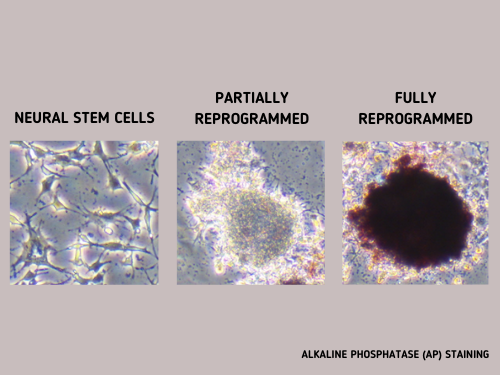
This photo shows cells screened using Alkaline Phosphatase (AP) staining. A group of scientists developed a new method that avoids invasive approaches that require stains or labels to track cell reprogramming status. Their novel technique uses “chemical fingerprint” data collected via non-invasive Raman spectroscopy to train an AI so it can monitor if the reprogramming of ordinary cells into stem cells is on track and check for markers that will verify a successful return to their earliest pluripotent stage.
The 2012 Nobel prize-winning discovery that ordinary cells could be coaxed to revert to their earliest pluripotent stage ushered in the era of ethical stem cell research. Suddenly, scientists can have an inexhaustible supply of pluripotent stem cells — the most versatile of stem cells — that can become any type of cell much like how embryonic stem cells function but without the ethical troubles that hampered research in the past.
These reprogrammed cells called induced pluripotent stem cells, or iPS cells, hold great promise for regenerative medicine, where they can be used to develop tissue or organ replacement-based treatments for life-threatening diseases.
Artificially inducing ordinary cells to reset back to pluripotency, however, is a lengthy and delicate process. Obtaining iPS cells depends on chance. And knowing all they can about the chemical changes cells undergo during reprogramming can help scientists improve the odds of attaining viable iPS cells for clinical applications. Current methods that track reprogramming status, however, use destructive and costly techniques.
A study led by Dr. Tomonobu Watanabe, professor at Hiroshima University’s Research Institute for Radiation Biology and Medicine, showed how Raman spectroscopy could be a low-cost, simpler, and non-intrusive technique in monitoring what goes on inside the cell as it transitions.
“The quality evaluation and sorting of existing cells have been carried out by investigating the presence or absence of expression of surface marker genes. However, since this method requires a fluorescent antibody, it is expensive and causes a problem of bringing the antibody into the cells,” Watanabe said.
“Solution of these problems can accelerate the spread of safe and low-cost regenerative medicine using artificial tissues. Through our method, we provide a technique for evaluating and sorting the quality of iPS cells inexpensively and safely, based on scattering spectroscopy,” he added.
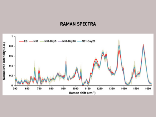
Raman spectroscopy avoids invasive approaches that require dyes or labels to extract biochemical information. Instead, it relies on vibration signatures produced when light beams interact with chemical bonds in the cell. As each chemical has a distinct vibration frequency, scientists can use it to identify the cell’s molecular composition.
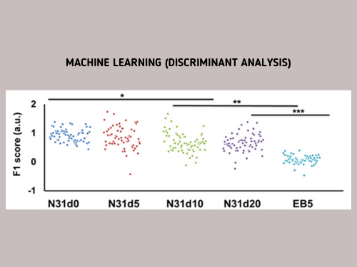
The team used this spectroscopic technique and followed mouse embryonic stem cells as they differentiated into neuronal cells then artificially reprogrammed back to pluripotency. They used the "chemical fingerprint" data they collected to teach an AI model so it can track if the reprogramming is progressing without a hitch and verify iPS cell quality by checking for a “fingerprint” match with the embryonic stem cell.
To measure the progress, they assigned the “chemical fingerprint” of neuronal cells as the transformation starting point and the embryonic stem cell’s patterns as the desired end goal. Along the axis, they used “fingerprint” samples collected on days 5, 10, and 20 of the neuronal cells’ reprogramming as reference points on how the process is advancing.
They published their findings in the October 2020 issue of the journal Analytical Chemistry.
“The Raman scattering spectrum contains comprehensive information on molecular vibrations, and the amount of information may be sufficient to define cells. If so, unlike gene profiling, it allows for a more expressive definition of cell function,” Watanabe said.
“We aim to study stem cells from a different perspective than traditional life sciences.”
(Research news authored by Mikas Matsuzawa)

 Home
Home