
 Home
HomeKey points of this research results
- Image enhancement using deep learning models trained on high-quality images enables significant noise reduction and improved spatial resolution in CT images.
- Lower noise in CT images allows for reduced radiation dose, making CT exams safer for patients.
- Improved spatial resolution allows for better visualization of fine structures, improving diagnostic accuracy.
Outline
CT imaging relies on differences in X-ray absorption within anatomical structures. However, image quality decreases under low radiation dose scans due to increased noise, which may reduce diagnostic performance. Hence, minimizing radiation exposure while maximizing diagnostic benefits remains a key challenge. With advances in deep learning, novel techniques have emerged for enhancing image quality, particularly in medical imaging(1). This study applies deep learning models to improve CT image quality. It is a collaborative effort between Hiroshima University and Canon Medical Systems Corporation.
The deep learning reconstruction method (DLR) we first developed is a technique that uses deep learning models trained with low-noise target images to generate high-quality images with reduced noise. As shown in Figure 1, it is now possible to obtain high-quality images by applying a deep learning model to cross-sectional images reconstructed using conventional methods from projection data obtained by CT scanning (2,3). The results shown in Figure 2 confirm that the noise is significantly reduced and higher-quality CT images are obtained compared to images obtained using conventional methods.
Next, we developed Super Resolution Deep Learning Reconstruction (SR-DLR), which enables the generation of low-noise, high-spatial-resolution images by training a deep learning model using target images with low noise and high spatial resolution(4,5). As shown in Figure 3, compared to images output by the conventional method, noise suppression and clear depiction of fine structures are possible, demonstrating the effectiveness of this method.
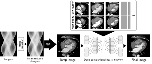
Fig. 1: Workflow showing the application of DLR and SR-DLR on CT data.
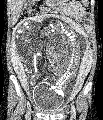
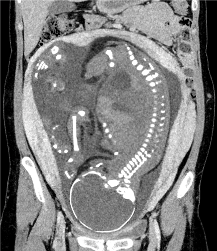
Fig. 2: Fetal CT image processed using DLR shows substantial noise reduction compared to the conventional method.

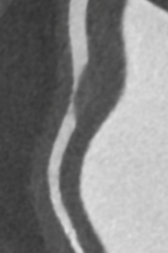
Fig. 3: Coronary artery phantom image processed using SR-DLR enhances fine structures that are unclear in conventional outputs.
Reference
- Higaki T, Nakamura Y, Tatsugami F, Nakaura T, Awai K. Improvement of image quality at CT and MRI using deep learning. Jpn J Radiol. 2019;37(1):73–80.
- Higaki T, Nakamura Y, Zhou J, et al. Deep learning reconstruction at CT: Phantom study of the image characteristics. Acad Radiol. Elsevier BV; 2020;27(1):82–87.
- Nakamura Y, Higaki T, Tatsugami F, Zhou J. Deep learning–based CT image reconstruction: initial evaluation targeting hypovascular hepatic metastases. Radiology: Artificial. pubs.rsna.org; 2019;
https://pubs.rsna.org/doi/abs/10.1148/ryai.2019180011. - Higaki T, Tatsugami F, Ohana M, Nakamura Y, Kawashita I, Awai K. Super resolution deep learning reconstruction for coronary CT angiography: A structured phantom study. Eur J Radiol Open. Elsevier BV; 2024;12(100570):100570.
- Tatsugami F, Higaki T, Kawashita I, et al. Improvement of Spatial Resolution on Coronary CT Angiography by Using Super-Resolution Deep Learning Reconstruction. Acad Radiol. 2023;30(11):2497–2504.